What is visual Dyslexia?
For normal vision to occur there should be binocular fixation in both eyes. However, many times the fixation of one macular is very unsteady, or there can be an eccentric fixation. This demonsrates the main cause for the learning problem, the patient is able to obtain a clear focus at any level of gaze but it cannot be maintained.
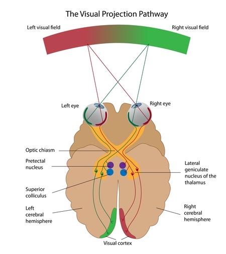
As fixation fails, so does the patients concentration. As there is unsteady or incorrect fixation, the frontal lobe of the brain lays down deep central suppression in the visual cortex (occipital cortex) of the affected eye.
This is what we term visual dyslexia; a physiological inability for inner vision resulting in a learning disability.
Visual Cortex
 The successful treatment of dyslexia is heavily involved with the visual cortex, which is at the back of the brain. In pure medical terms, it is situated on the medial aspect of the occipital lobe in relation to the calcarine fissure. It is characterised by the distinguishing white line or stria of Gennari, which is visible to the naked eye. The cellular structure of the visual cortex is of the highly granular type associated elsewhere in the cortex with sensory function. The outer and inner granular layers are made up of small granular cells densely packed.
A simple analogy of the visual cortex likens it to that of a Layer cake with seven layers, the eye simply being the camera. Utilising the LASD (Lawson Anti-Suppression Device), the larger discs stimulate the upper layers and the finest bands stimulating the deepest layer of the visual cortex.
The eye is like the camera: The visual cortex in the occipital (lower back part of the brain) area is the main receiving station of the visual impulses, and the frontal lobe (upper area) of the brain interprets what is being seen.
